มาทำคลอดฟันเขี้ยวฝัง ออกมาสู่ช่องปากกันเถอะ!
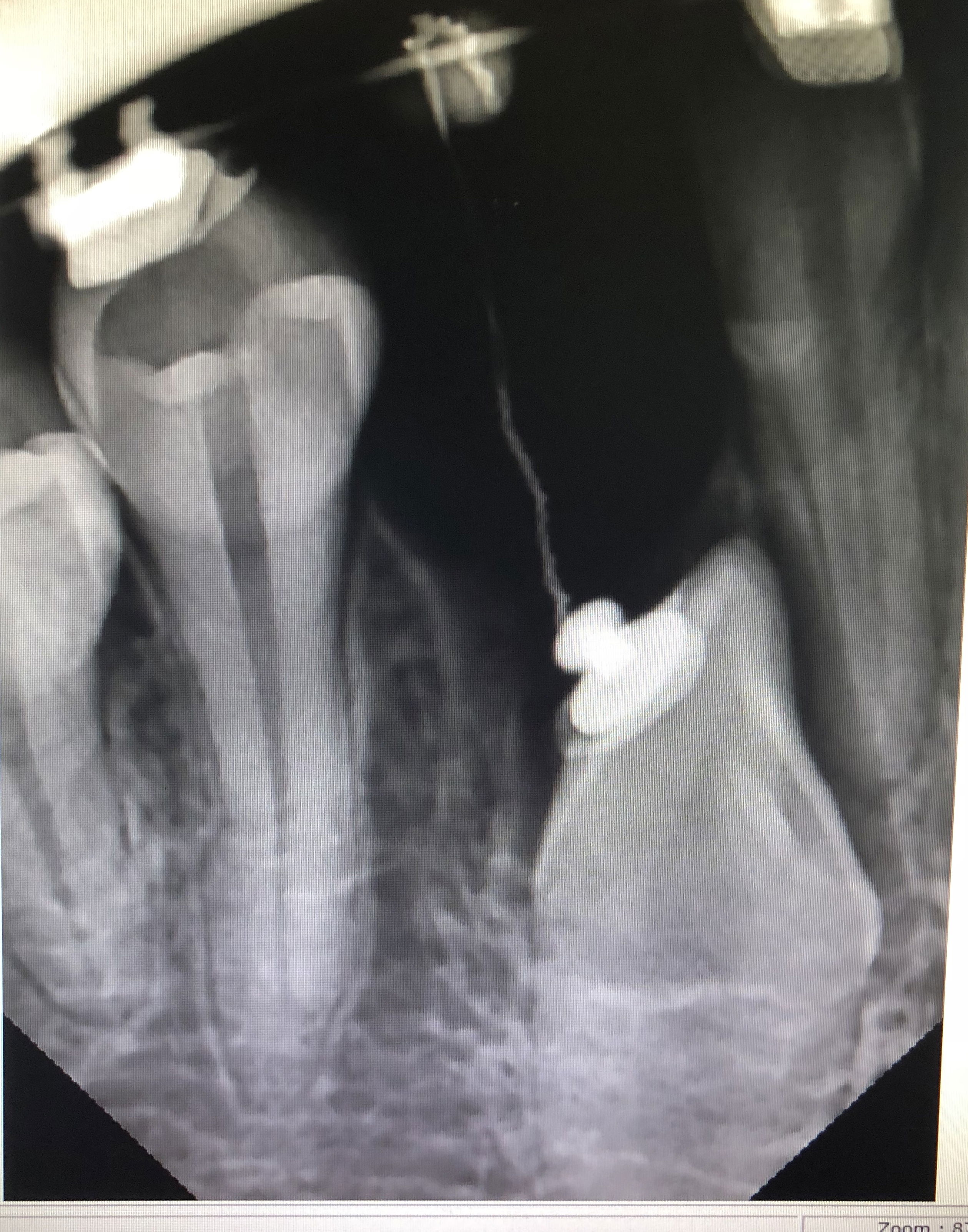
รีวิวเคสตัวอย่าง ของคลินิกทันตกรรมเดอะคราวน์ จากภาพถ่ายรังสีเอ็กซ์เรย์ พบฟันเขี้ยวล่างด้านขวาฝังในกระดูกขากรรไกร วางในลักษณะตรง และมีฟันน้ำนมคงเหลือในช่องปาก คุณหมอจึงพิจารณาถอนฟันน้ำนมออก และวางแผนนำฟันเขี้ยวซี่นี้ออกมาในช่องปาก ซึ่งเมื่อถอนฟันน้ำนมออกมา พบว่า ยังมีกระดูกคลุมตัวฟันเขี้ยวอยู่ จึงพิจารณาศัลยกรรม กรอแต่งกระดูกที่ปกคลุมฟันออก ให้สามารถติดเครื่องมือจัดฟันบริเวณตัวฟันเขี้ยวได้

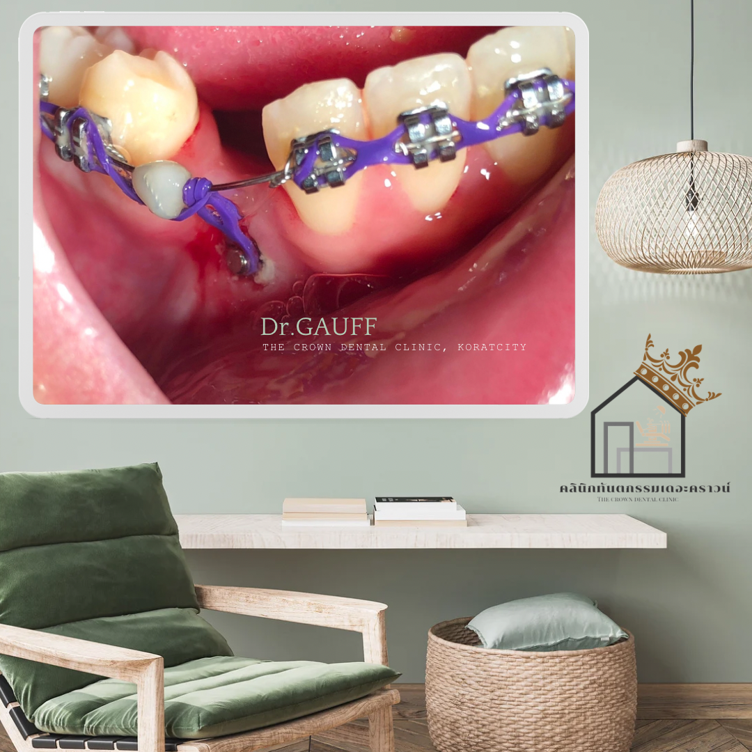
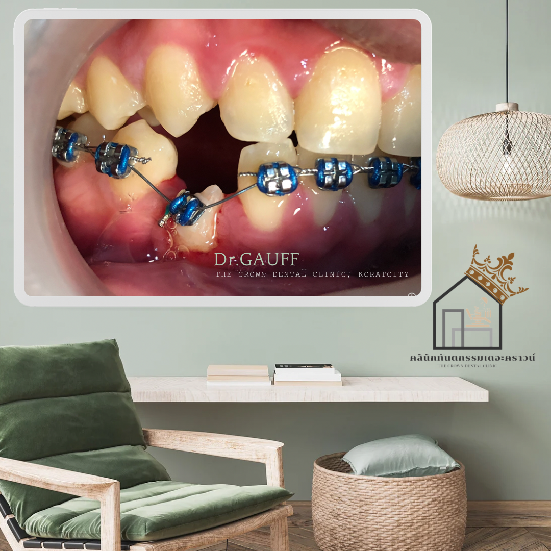
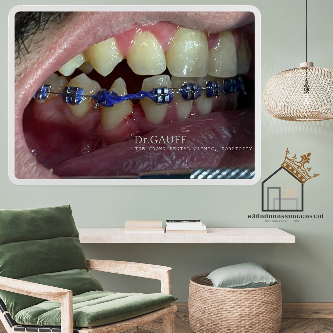
ณ ปัจจุบัน คนไข้รายนี้ ยังอยู่ในขั้นตอนของการปิดช่องห่างของฟันล่าง และคุณหมอทดสอบความมีชีวิตของฟันซี่นี้ พบว่าฟันซี่นี้ให้ผล + ต่อเครื่องทดสอบความมีชีวิตของฟัน พบร่องลึกปริทันต์ ประมาณ 4 มม.

Sample Case Review – The Crown Dental Clinic, KoratRadiographic examination revealed an impacted lower right canine embedded within the jawbone, positioned in an upright orientation. A retained primary (deciduous) tooth was also present in the oral cavity. The dentist therefore decided to extract the primary tooth and plan for guided eruption of the impacted canine into the oral cavity.After removal of the primary tooth, it was found that the impacted canine was still covered by bone. As a result, surgical exposure and bone contouring were performed to remove the overlying bone, allowing orthodontic appliances to be bonded to the canine for orthodontic traction.At present, the patient is still in the phase of closing the space in the lower arch. Pulp vitality testing of the impacted canine showed a positive response, indicating that the tooth remains vital. Periodontal probing revealed a probing depth of approximately 4 mm around the tooth.
ขอบคุณที่ติดตามอ่านบทความ : คุณหมอกอล์ฟ คลินิกทันตกรรมเดอะคราวน์โคราช