รีวิว เคสรากเทียมฟันหน้า 2 ซี่ ผ่านทันตนวัตกรรมปัจจุบัน
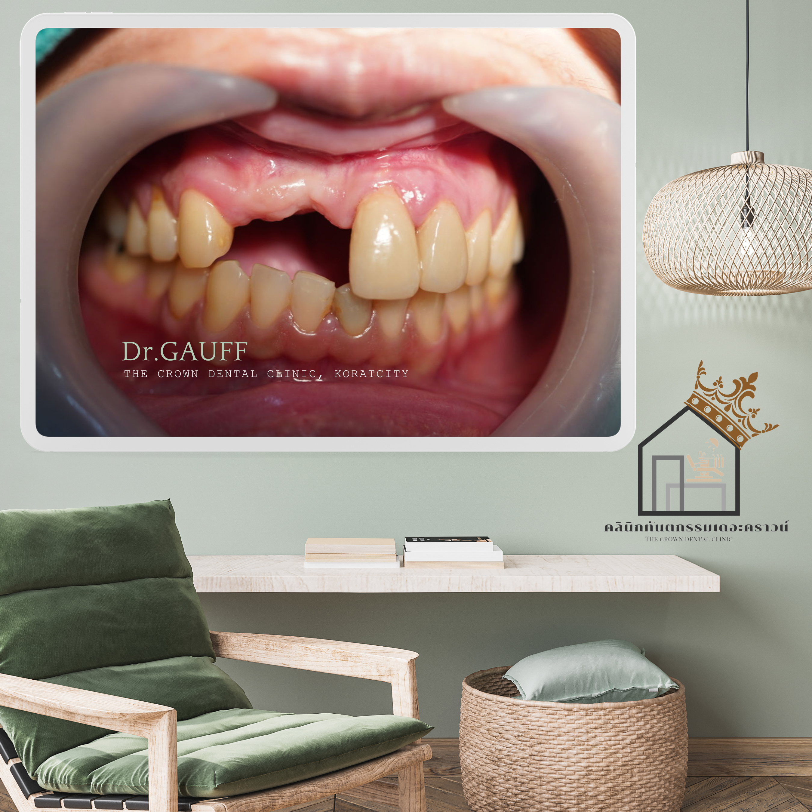
เคสคนไข้ของคลินิก ต้องการทดแทนฟันหน้า 2 ซี่ ด้วยรากฟันเทียม
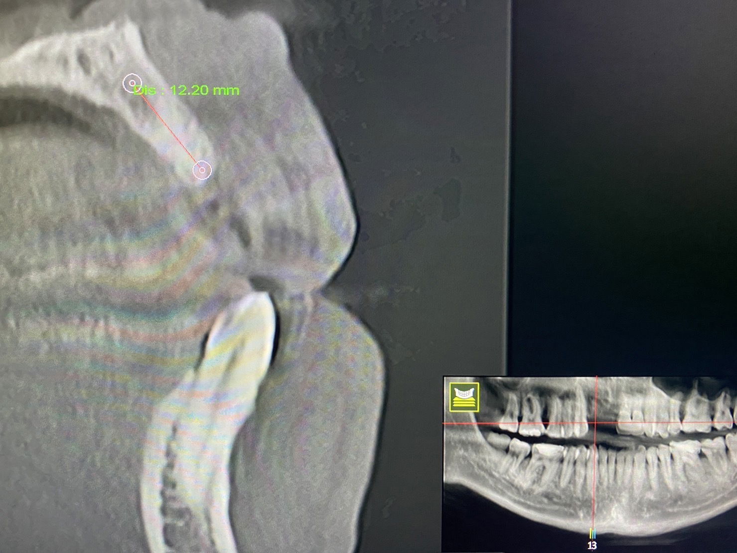
คลินิกทันตกรรมเดอะคราวน์ ใช้เครื่อง 3D X-Ray หรือเครื่อง CT Scan เพื่อให้ได้ภาพของกระดูกขากรรไกร แบบสามมิติ เพื่อนำมาวางแผนการรักษาฝังรากเทียมได้อย่างแม่นยำมากขึ้น

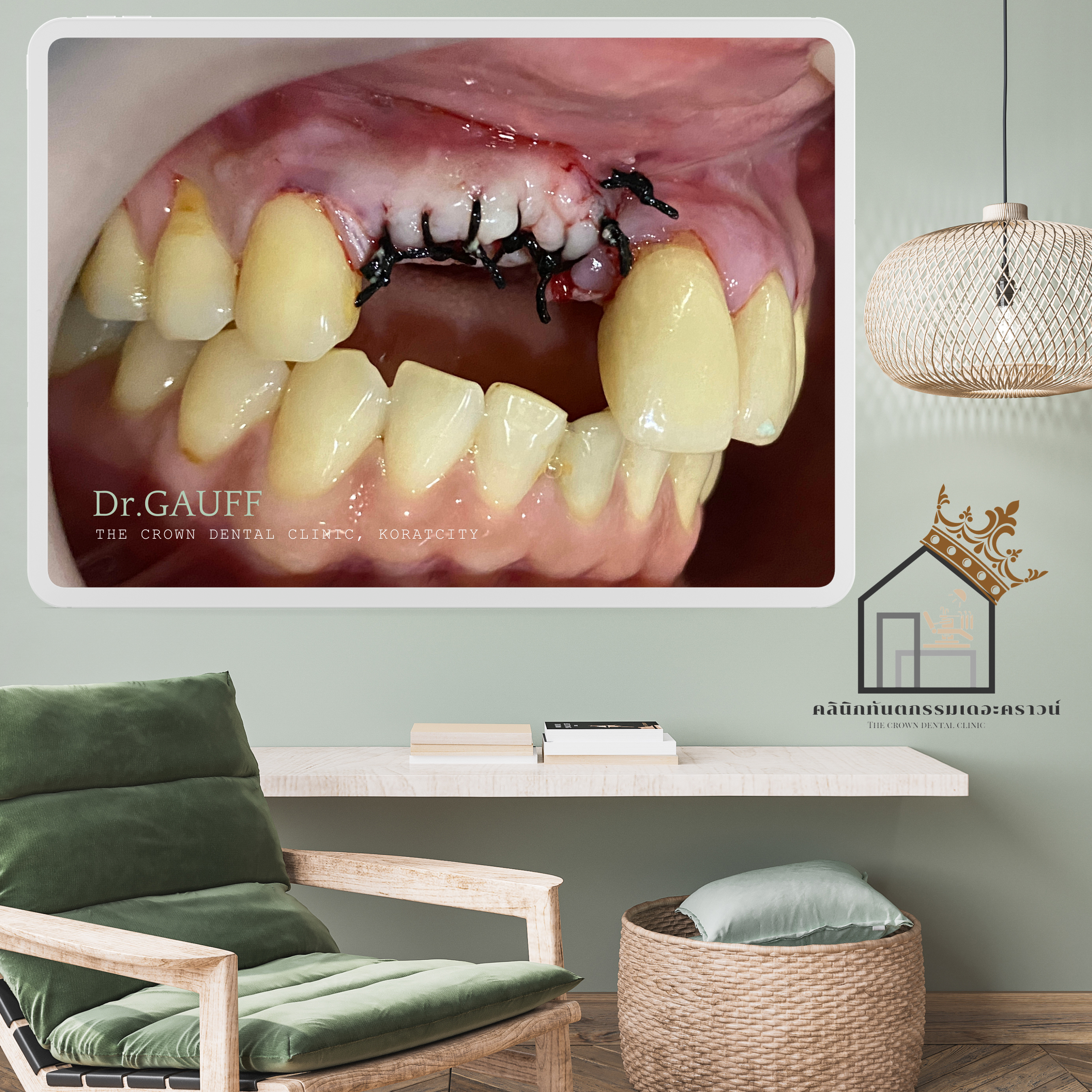
ในขั้นตอนของการฝังรากเทียม คุณหมอวางแผนการรักษาในการปลูกกระดูกเทียม ร่วมกับเหงือกเทียม เพื่อทดแทนกระดูกบริเวณฟันหน้าที่สูญเสียไปจากการถอนฟัน เนื่องจากฟันหน้าเป็นตำแหน่งที่เกี่ยวข้องกับความสวยงามอย่างมาก และการฝังรากเทียมในแต่ละครั้งมักมีการละลายของกระดูกในบริเวณนี้เพิ่ม ดังนั้นการปลูกกระดูกในบริเวณดังกล่าว จึงช่วยชะลออัตราการการละลายของกระดูกด้วย
หลังจากการรอระยะของการหายของแผล ระยะการสร้างกระดูก และการเกิดการยึดของกระดูกเข้ากับส่วนของรากเทียม ประมาณ 5-6 เดือน ก็จะเข้าสู่ขั้นตอนที่ 2 ของการรักษา คือการทำส่วนของครอบฟันต่อไป
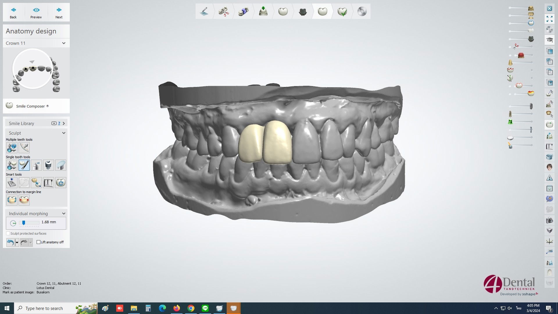
ในขั้นตอนการพิมพ์ปาก ในปัจจุบัน มีนวัตกรรมทางทันตกรรมที่เข้ามาช่วยให้การพิมพ์ปากง่ายมากขึ้น แม่นยำมากขึ้น ด้วยเครื่องสแกนฟัน ซึ่งช่วยลดขั้นตอนที่ยุ่งยาก และมีความผิดพลาดสูงได้มาก คนไข้รู้สึกสบายมากขึ้นอีกด้วย จากนั้น คุณหมอจะนำข้อมูลทั้งหมดส่งให้ทางห้องแล็บ ได้ทำการออกแบบส่วนของครอบฟัน ด้วยโปรแกรมคอมพิวเตอร์ดีไซน์ และเทคโนโลยีคอมพิวเตอร์ในการผลิตงาน (CAD/CAM)
โดยในขั้นตอนนี้ ปัจจุบันมีเทคโนโลยีการออกแบบชิ้นงาน ผ่านโปรแกรมคอมพิวเตอร์ ทำให้ส่วนของครอบฟันที่สร้างขึ้น มีความแม่นยำกับตำแหน่งจริงมากที่สุด
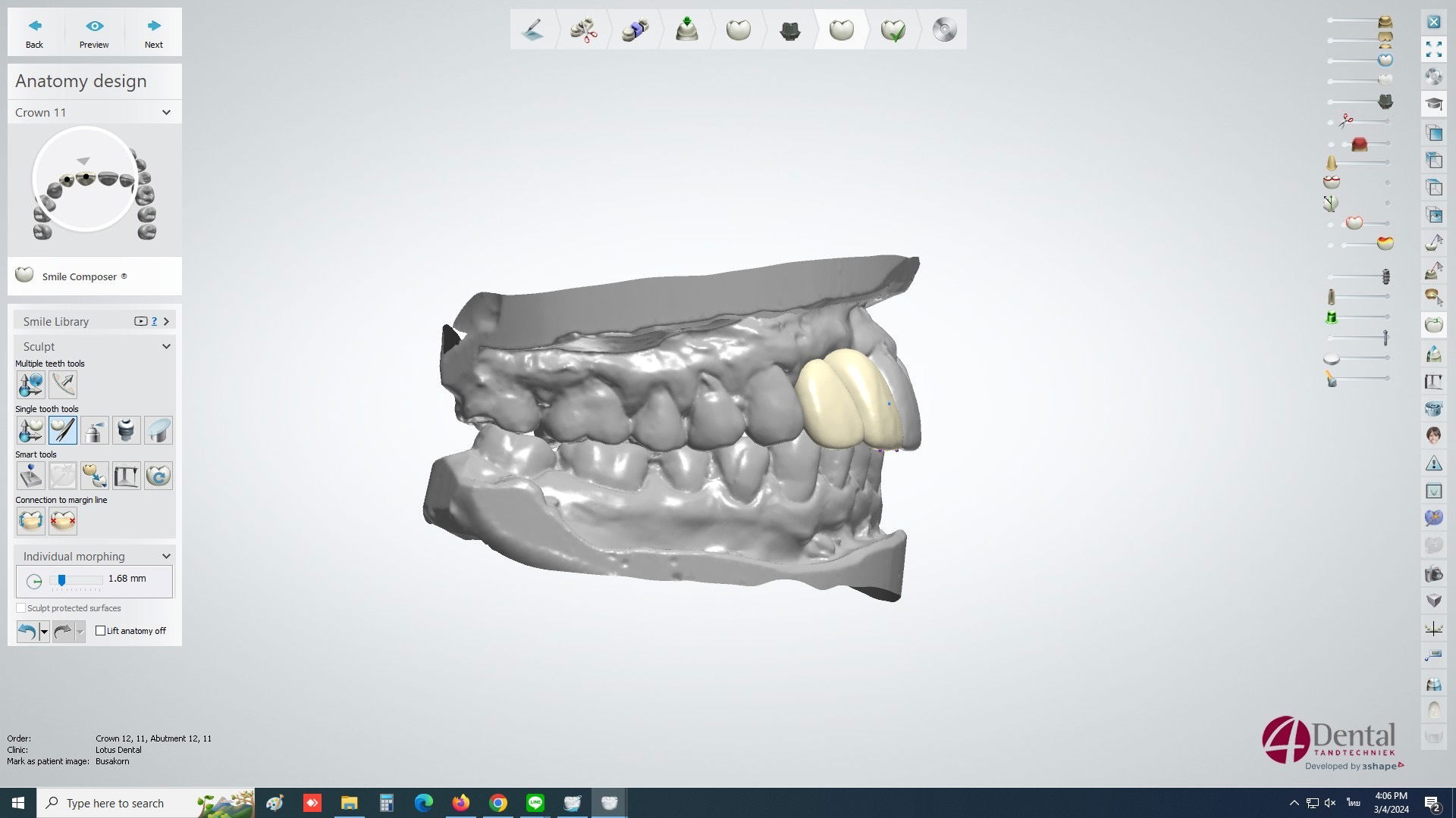
ขั้นตอนของการใส่ - จากการใช้เทคโนโลยี และนวัตกรรมทางทันตกรรม ณ ปัจจุบัน มาช่วยในการทำงาน ส่งผลให้ชิ้นงานที่นำมาใส่ในช่องปากให้กับคนไข้ มีความแม่นยำอย่างมาก ได้ในตำแหน่งที่เหมาะสม ตามที่ได้ออกแบบไว้ในโปรแกรมคอมพิวเตอร์
Patient Case Review – The Crown Dental Clinic, Korat
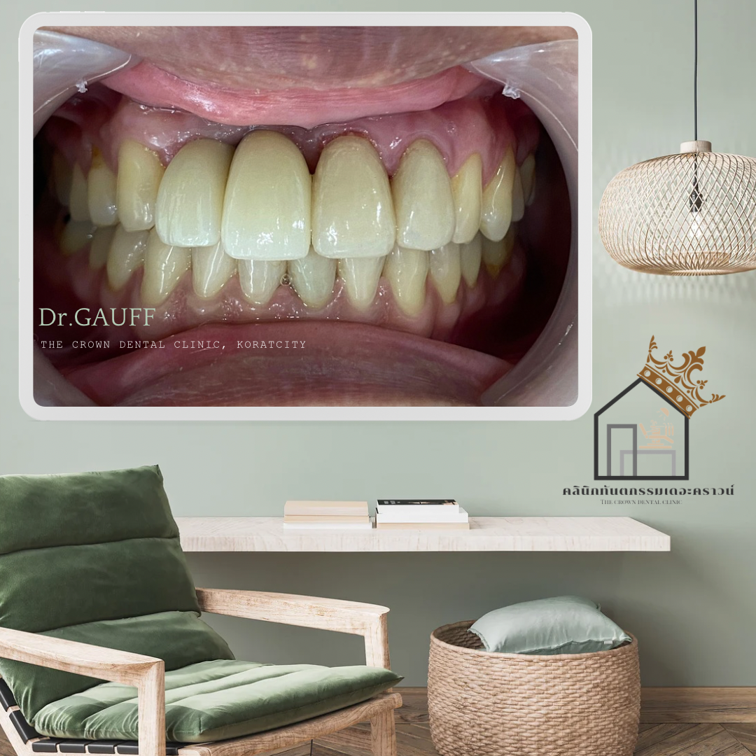
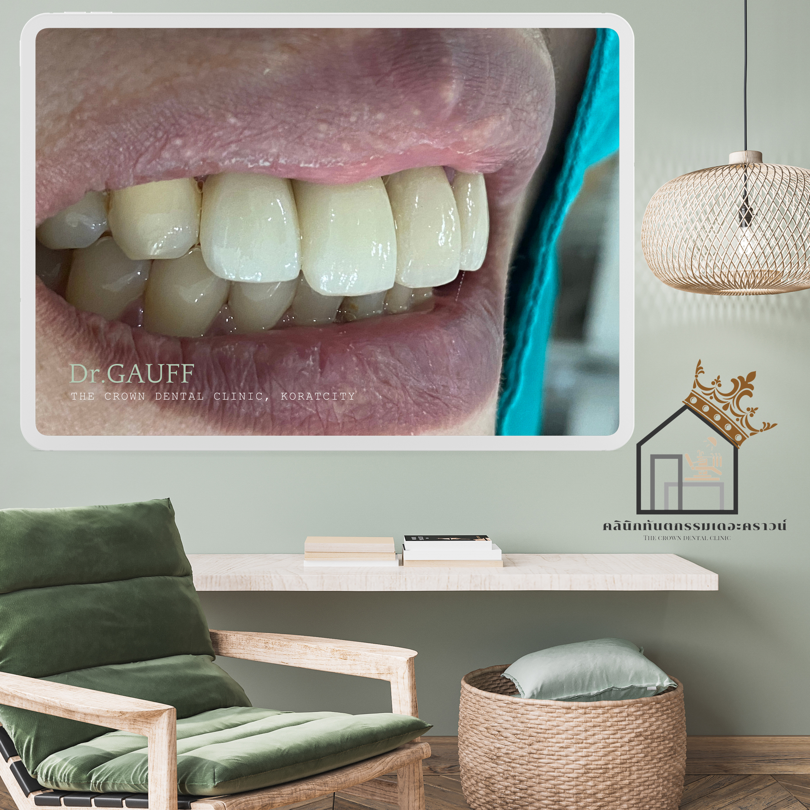
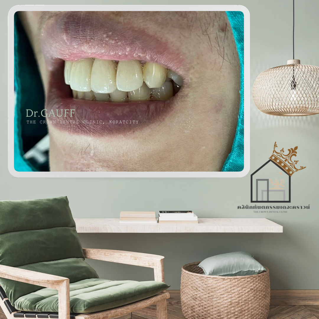
Replacement of Two Anterior Teeth with Dental ImplantsThe patient visited the clinic seeking replacement of two missing front teeth with dental implants.At The Crown Dental Clinic, a 3D X-ray (CT scan) is used to obtain three-dimensional images of the jawbone. This allows for highly accurate implant treatment planning, enabling precise evaluation of bone volume, bone quality, and the anatomical structures involved.
Implant Planning and Bone Grafting
During the implant placement phase, the dentist planned bone grafting together with soft tissue (gum) augmentation. This was necessary to compensate for bone loss in the anterior region following tooth extraction.
The front teeth are critically important for esthetics, and implant placement in this area is often associated with further bone resorption. Therefore, bone grafting in this region helps reduce the rate of bone loss and supports long-term esthetic outcomes.
Healing Phase and Second Stage Surgery
After a healing period of approximately 5–6 months, allowing for wound healing, new bone formation, and osseointegration (bone bonding to the implant surface), treatment progressed to Stage 2, which involved fabrication of the final crowns.
Digital Impression and CAD/CAM Technology
In the impression stage, modern dental innovations were utilized to improve accuracy and patient comfort. Intraoral digital scanning was used instead of conventional impression materials, significantly reducing complexity and potential errors. This method is more comfortable for patients and provides highly precise digital data.The collected data were then sent to the dental laboratory, where the crowns were designed using computer-aided design (CAD) software and fabricated using computer-aided manufacturing (CAM) technology.With current digital design technologies, the crowns can be fabricated with exceptional precision, closely matching the actual implant positions as planned virtually.
Crown Placement
Thanks to the integration of advanced dental technologies and digital workflows, the final restorations placed in the patient’s mouth achieved high accuracy, excellent fit, and optimal positioning, precisely as designed in the computer planning stage. This approach enhances both function and esthetics, especially in the anterior region where precision is essential.

ขอบคุณที่ติดตามอ่านบทความ : คุณหมอกอล์ฟ คลินิกทันตกรรมเดอะคราวน์โคราช