เคสงานแก้ รื้อสะพานฟัน ปิดจ็อบวีเนียร์เซรามิก
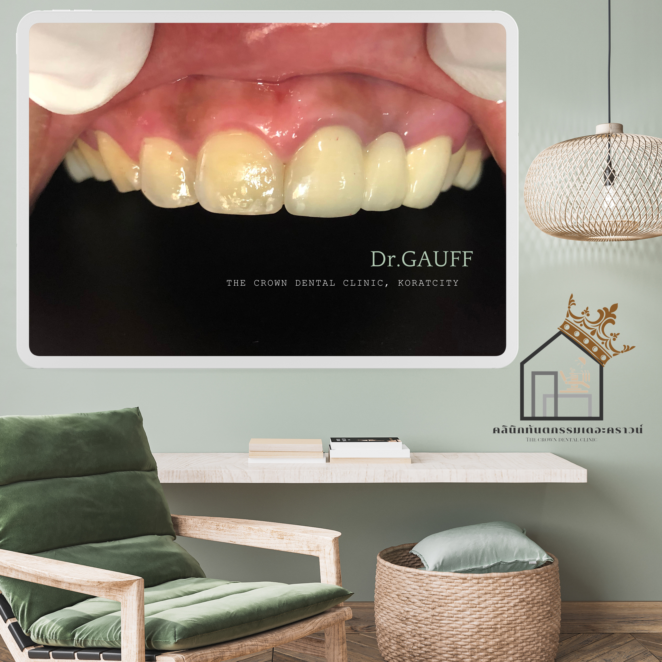
รีวิวเคสสมัย 6-7 ปีที่แล้ว ของคลินิกทันตกรรมเดอะคราวน์ เคสนี้อยากขอแก้ไขสีฟันหน้าให้ขาวขึ้น และยาวมากขึ้น โดยมีการบูรณะสะพานฟันด้านซ้ายมา และมีฟันกรามน้อยด้านเดียวกันนี้ผุทะลุโพรงประสาทฟัน
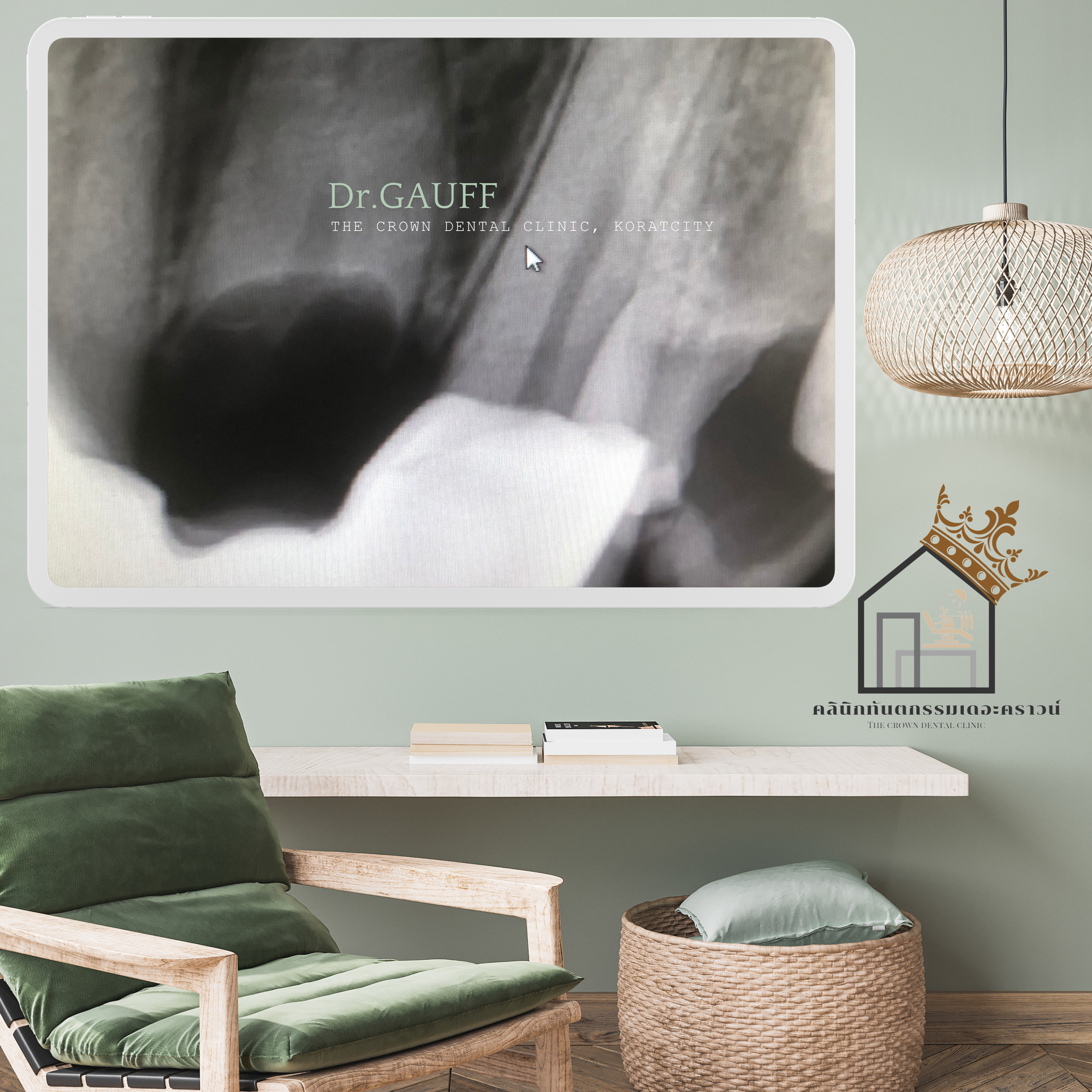
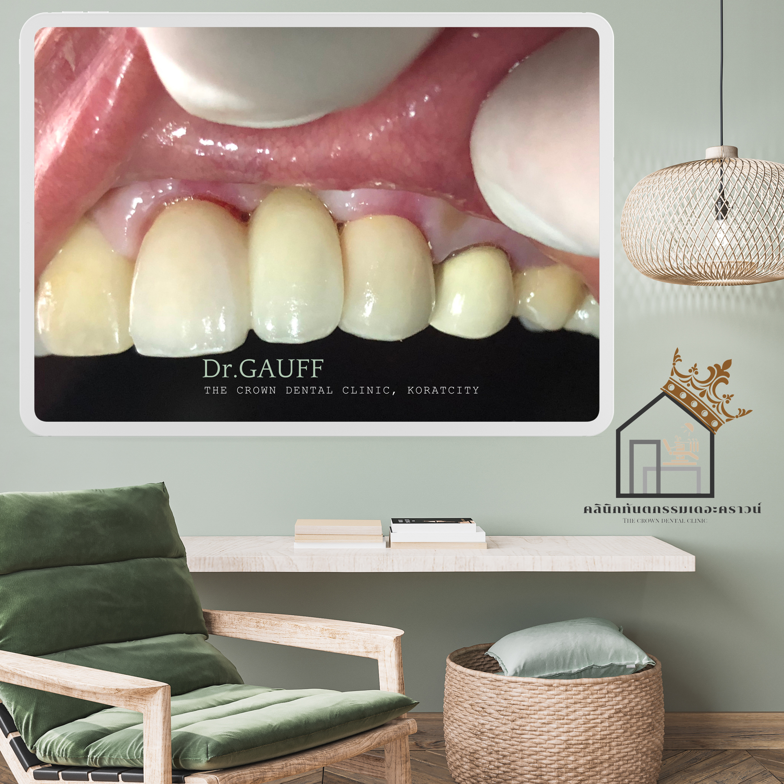
ส่ง X-Ray บริเวณสะพานฟันที่ทำมา พบว่า มีรากฟันเหลืออยู่ทั้งราก ใต้สะพานฟัน จึงแนะนำคนไข้ว่า ต้องรื้อสะพานฟันออก เพื่อผ่าเอารากฟันซี่นี้ออก ตำแหน่งของฟันเมื่อยิ้มแบบ extremely Smile พบว่าสามารถศัลยกรรมปริทันต์เพิ่มความยาวตัวฟันได้
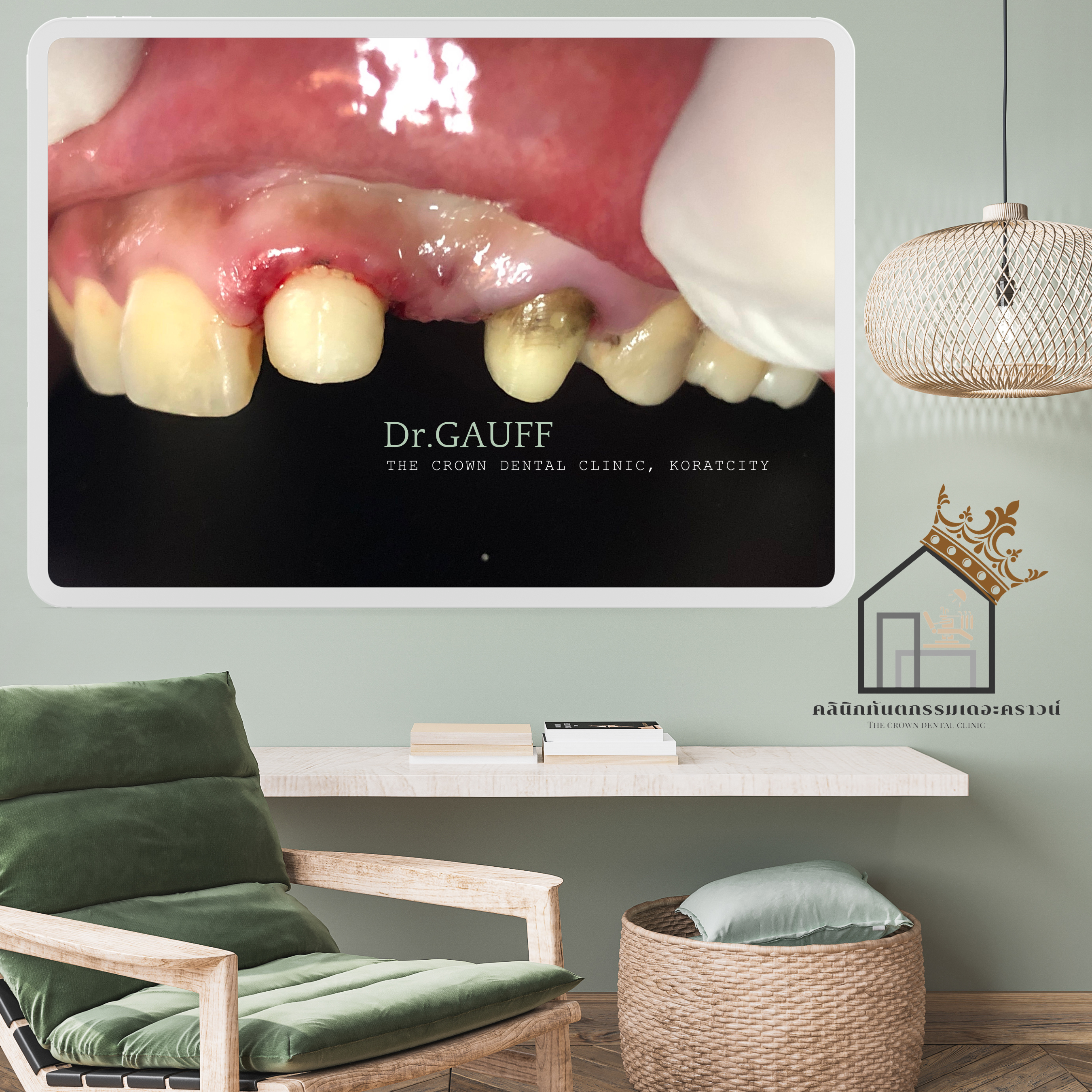
แนะ รื้อสะพานฟัน ผ่ารากฟันออก แต่งเหงือกให้ได้ระดับประมาณริมฝีปากบน ณ ตำแหน่ง extremely smile แล้วทำส่วนของสะพานฟันชั่วคราว ระหว่างรอ healing ก็รักษารากฟันกรามน้อยไปพร้อม ส่วนด้านขวาวางแผนทำงานวีเนียร์เซรากมิก

ในขั้นตอนของการบูรณะด้วยสะพานฟัน ครอบฟันกรามน้อย (ด้านซ้าย) และวีเนียร์(ด้านขวา) ควรทำพร้อมกันในครั้งเดียว เนื่องจากช่างทันตกรรมจะได้ทำงานออกมาครั้งเดียว ได้สี และรูปร่างลักษณะที่ค่อนข้างเหมือนกัน (ช่างสมัยเมื่อ 6-7 ปีก่อน ยังไม่ได้ใช้คอมพิวเตอร์ดีไซด์)
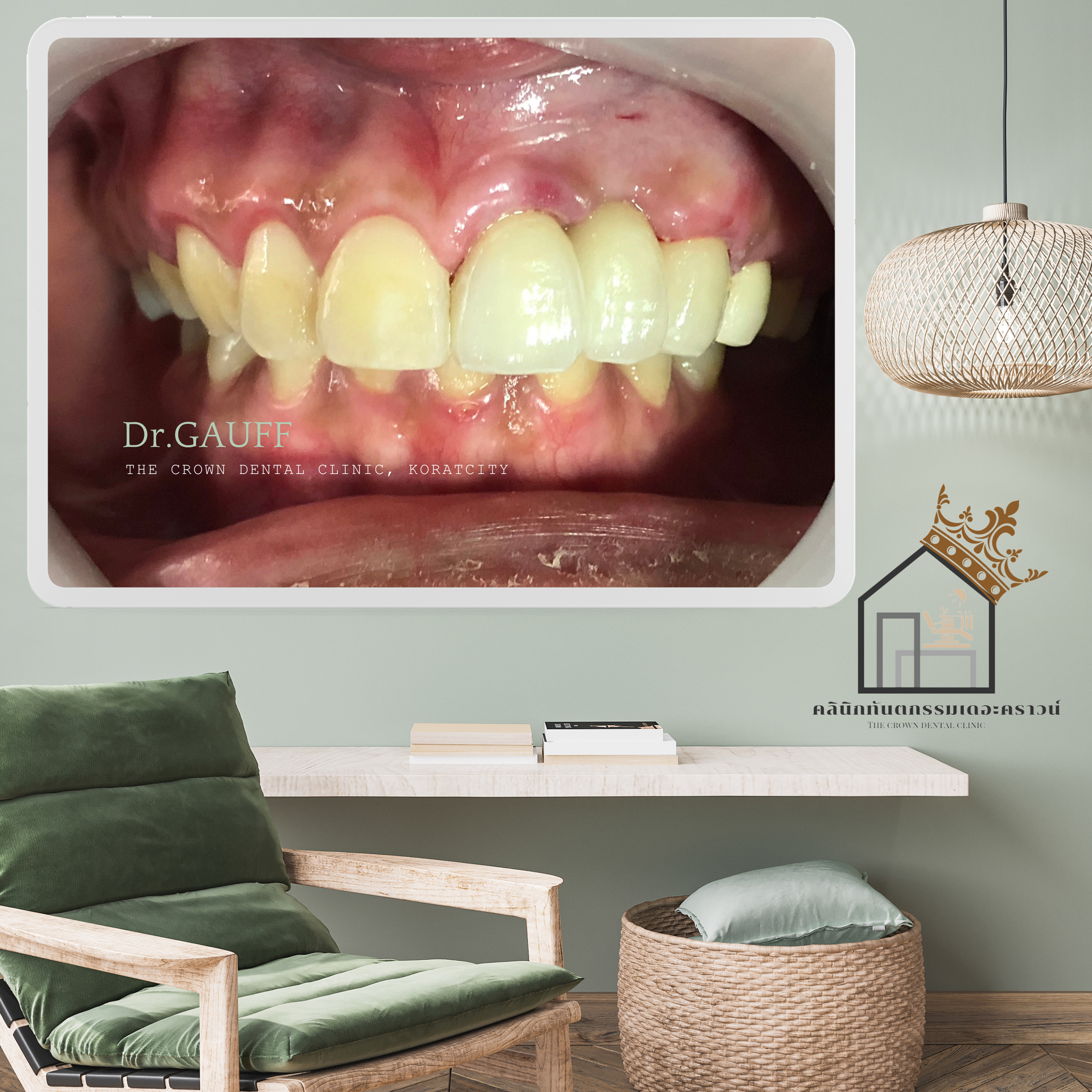
ด้านซ้าย (ของคนไข้ ) บูรณะด้วยสะพานฟันกลับไปเหมือนเดิม โดยให้คำแนะนำเรื่องการทำความสะอาดบริเวณสะพานฟันให้ดี และกลับมาติดตามเป็นระยะ เนื่องจากทำความสะอาดได้ค่อนข้างยาก อาจเกิดฟันผุหรือโรคเหงือกได้ง่าย หากเกิดฟันผุที่ฟันหลัก จะแก้ไขได้ยาก (ต้องรื้อสะพานออก เพื่ออุดฟัน แล้วทำสะพานฟันชุดใหม่กลับไป)
ส่วนทางด้านขวา(ของคนไข้) บูรณะด้วยงานวีเนียร์เซรามิก จำนวน 3 ซี่
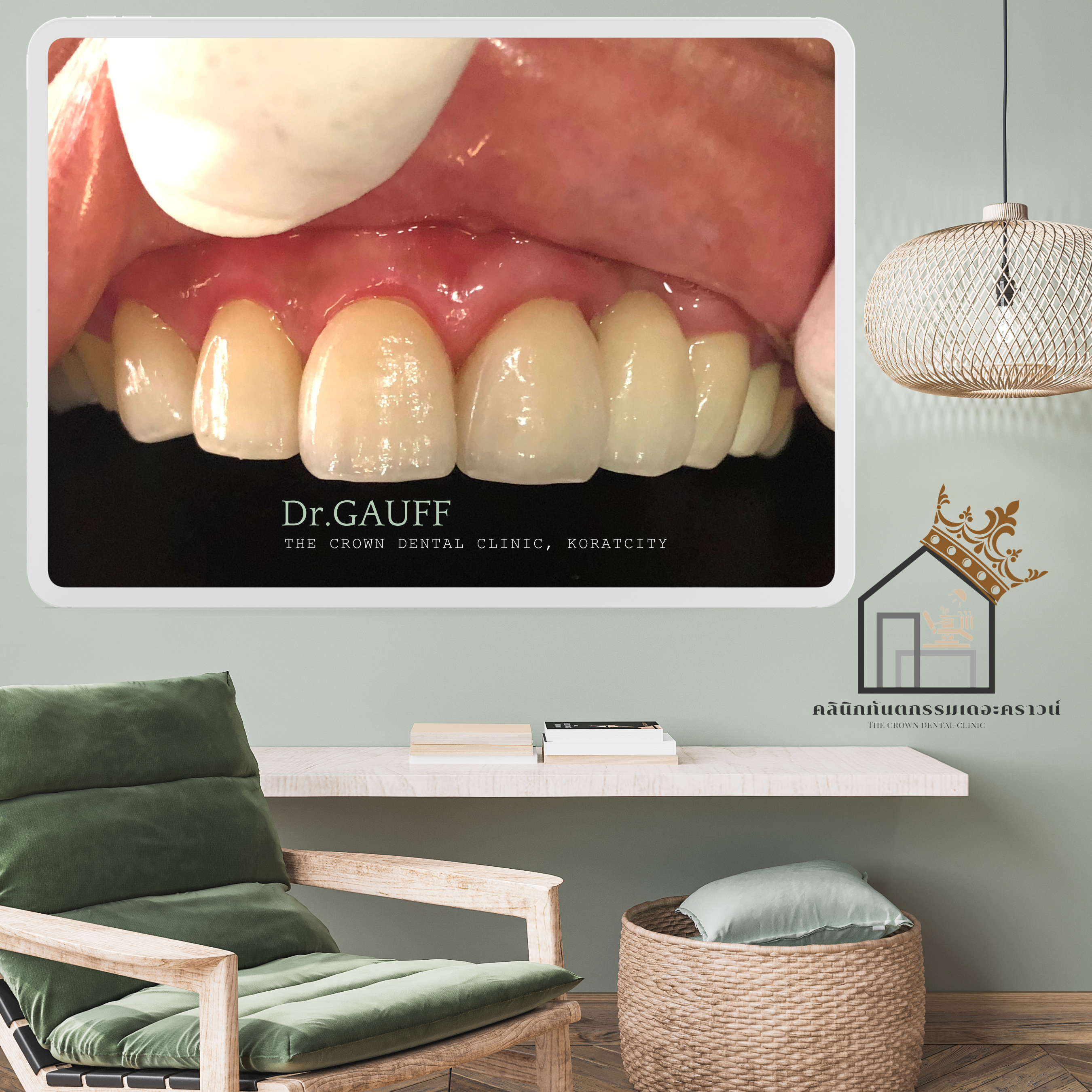
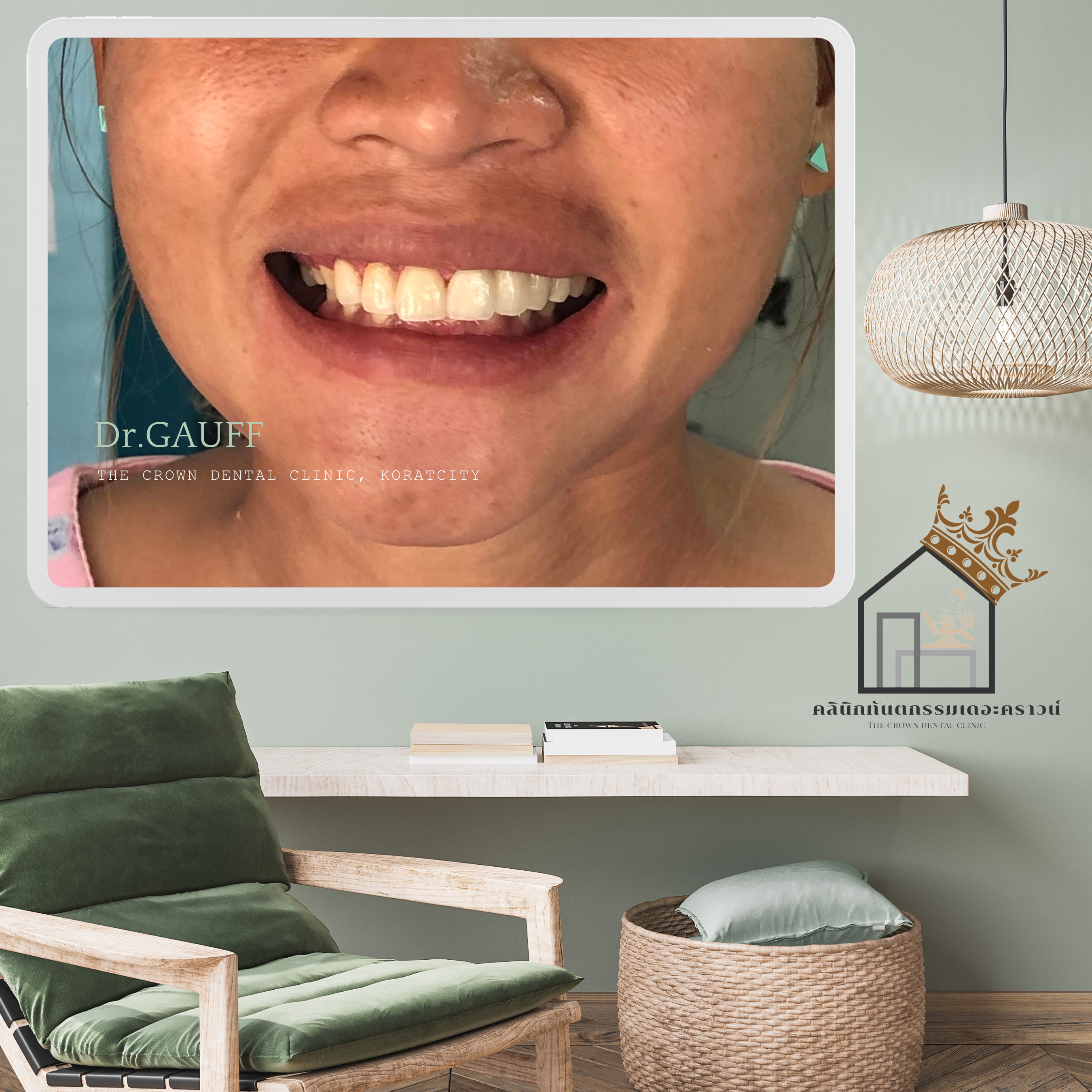
Case Review from 6–7 Years Ago – The Crown Dental ClinicIn this case, the patient wished to improve the color and length of the anterior teeth. The patient had an existing fixed bridge on the left side, and a premolar on the same side with caries extending into the dental pulp.Radiographic examination of the bridge area revealed that a retained root fragment remained completely under the bridge. The patient was therefore advised that the bridge needed to be removed in order to surgically extract the remaining root. When evaluating the smile in an extreme smile position, it was determined that periodontal surgery (crown lengthening) could be performed to increase the visible length of the teeth.The treatment plan included removal of the existing bridge, surgical extraction of the retained root, and gingival recontouring to a level appropriate for the upper lip during an extreme smile. A temporary bridge was fabricated during the healing period. At the same time, root canal treatment of the premolar was carried out. On the right side, ceramic veneer restorations were planned.For the definitive restorations—including the bridge and premolar crown on the left side and the veneers on the right side—it was recommended that all restorations be fabricated at the same time. This allowed the dental technician to produce the work in a single stage, ensuring better consistency in shade and morphology, especially since computer-aided design (CAD) technology was not yet widely used 6–7 years ago.On the left side, the bridge was restored in a form similar to the original design. The patient was advised on meticulous oral hygiene around the bridge area and regular follow-up visits, as cleaning around bridges can be challenging and may increase the risk of caries or periodontal disease. If decay were to occur on an abutment tooth, management would be complex, often requiring removal of the bridge, treatment of the tooth, and fabrication of a new bridge.On the right side, the patient was restored with three ceramic veneers.
ขอบคุณที่ติดตามอ่านบทความ คุณหมอกอล์ฟ คลินิกทันตกรรมเดอะคราวน์โคราช