เคสที่มีฟันฝัง ฟันคุด ต้องมาฟัง!! การเรียงตัวของซี่ฟันบางซี่ผิดปกติ มักมีรอยโรครุนแรงซ่อนอยู่?
รีวิวเคสของคลินิกทันตกรรมเดอะคราวน์ ตรวจพบรอยโรคที่มีระดับรุนแรง ซึ่งสัมพันธ์กับฟันคุด หรือฟันฝัง ที่ยังไม่ได้รับการนำออกมา
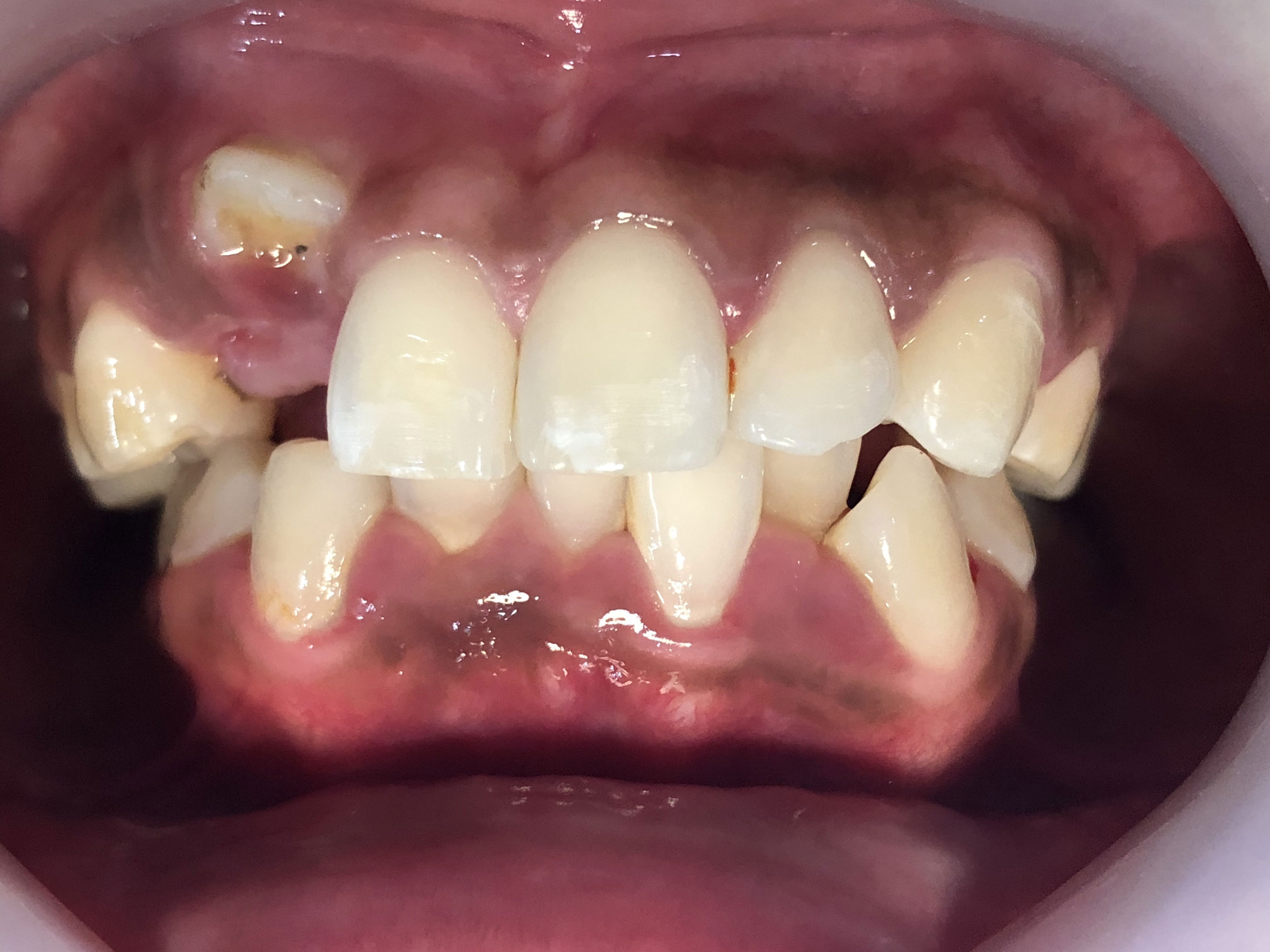
เคสแรก เป็นเคสจัดฟัน มีฟันหน้า ขึ้นในตำแหน่งที่ผิดปกติ ภาพถ่ายเอ็กซ์เรย์บริเวณดังกล่าวพบ ฟันเขี้ยวฝัง และมีถุงน้ำ (Cyst) แนะนำผ่าเอาฟันฝังออก และฟันหน้าซี่ดังกล่าวต้องนำออกด้วย เพราะ Cyst จะทำลายกระดูโดยรอบ ซึ่งหากปล่อยไว้ Cyst อาจจะละลายกระดูกที่รอบรากฟันของซี่ที่ขึ้นปกติ ทำให้ฟันซี่ปกตินั้น เกิดฟันโยกในที่สุด
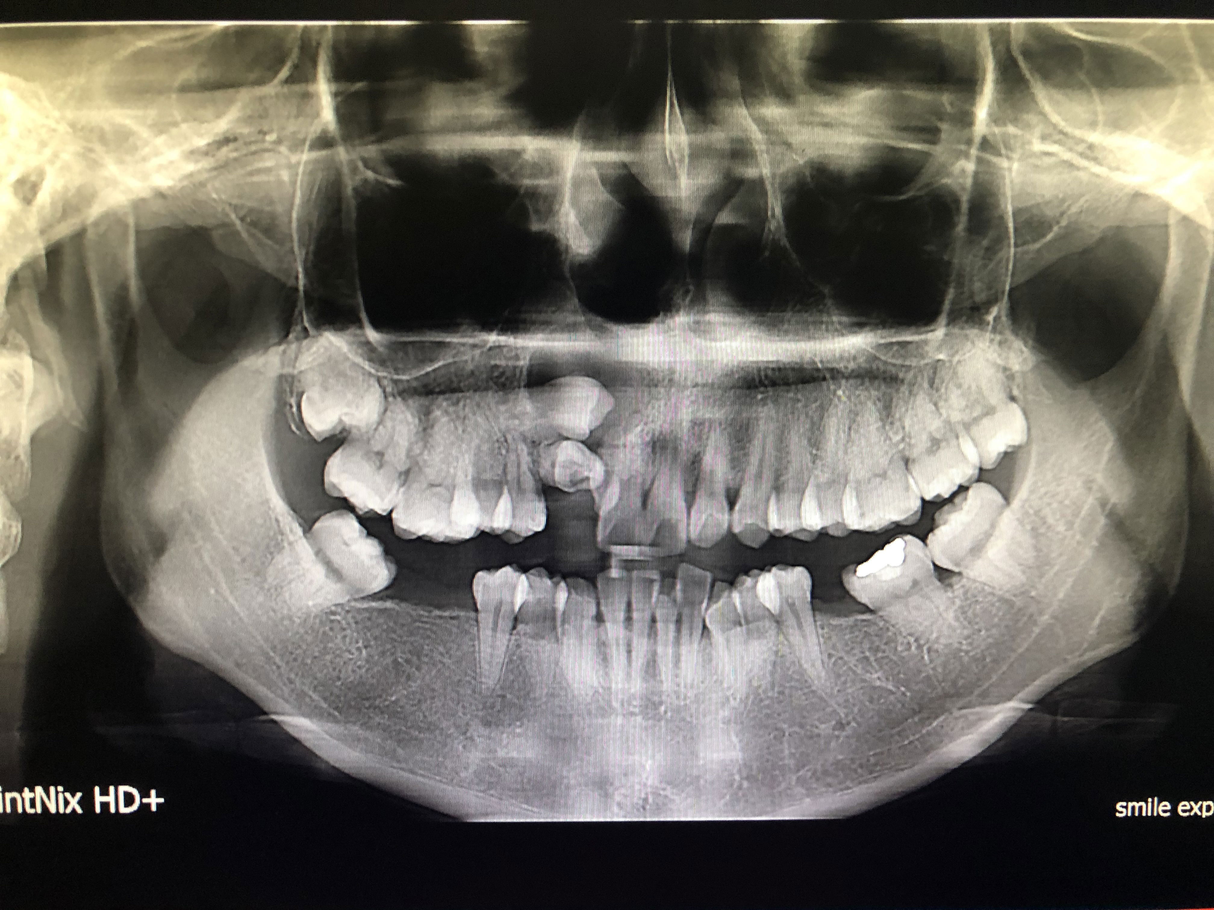
จากภาพถ่ายรังสีเอ็กซ์เรย์ ด้านขวาของภาพ (ด้านซ้ายของคนไข้) บริเวณฟันคุดล่างด้านซ้าย จะพบลูกศรสีขาว เป็นก้อนมีขอบเขตชัดเจนสัมพันธ์กับฟันคุด รอยโรคนี้คุณหมอส่งเอ็กซ์เรย์เป็นเคสคนไข้จัดฟัน เนื่องจากคนไข้ให้ประวัติปวดฟันคุด มีน้ำเค็มๆไหลออกมา และหน้าบวม
รอยโรคดังกล่าวจากการส่งตัวคนไข้ ไปพบคุณหมอศัยกรรมช่องปาก พบว่า เป็นเนื้องอกกรามช้าง หรือ Ameloblastoma เป็นเนื้องอกชนิดที่ไม่รุนแรง ที่มีจุดกำเนิดมาจากฟัน และจากสถิติพบมากที่สุดในประเทศไทย
โดยจะมีการบวมโตของขากรรไกรอย่างช้าๆ ไม่ค่อยมีอาการเจ็บปวดใดๆ ถึงแม้ว่าเนื้องอกกรามช้างจะไม่ใช่มะเร็ง แต่มีพฤติกรรมค่อนข้างรุนแรง มีการทำลายกระดูกขากรรไกรรอบๆรอยโรคได้มาก พบบ่อยที่ขากรรไกรล่าง
การรักษาคือการผ่าตัดเอารอยโรคและกระดูกรอบรอยโรคออก หากเกิดการทำลายอย่างมาก อาจจะต้องตัดขากรรไกรออกด้วย เนื่องจากเนื้องอกชนิดนี้สามารถกลับมาเป็นซ้ำได้สูง ดังนั้นจึงต้องมีการตรวจติดตามตามระยะ
หากท่านมีฟันคุด หรือมีฟันฝัง ที่ได้รับการตรวจยืนยันทางภาพถ่ายรังสี จากทันตแพทย์แล้ว ท่านควรพิจารณาผ่าตัดเอาออกตั้งแต่เริ่มแรก จะช่วยลดโอกาสการเกิดถุงน้ำ หรือแม้กระทั่งเนื้องอกชนิดต่างๆได้
Case Review – The Crown Dental Clinic, Korat: Severe Pathologic Lesions Associated with Impacted TeethIn this review, severe pathologic lesions were detected in association with impacted or unerupted teeth that had not yet been removed.
Case 1
This was an orthodontic case in which an anterior tooth erupted in an abnormal position. Radiographic examination revealed an impacted canine associated with a cystic lesion. Surgical removal of the impacted tooth was recommended, and the affected anterior tooth also needed to be extracted. This was because the cyst had the potential to destroy the surrounding bone. If left untreated, the cyst could resorb the bone around the roots of adjacent normally erupted teeth, eventually leading to tooth mobility of those teeth.
Case 2
On the radiographic image, on the right side of the image (corresponding to the patient’s left side), a well-defined radiolucent lesion can be seen adjacent to the lower left impacted wisdom tooth, indicated by a white arrow. This case was referred as part of an orthodontic evaluation. The patient reported symptoms of pain in the wisdom tooth area, discharge of salty-tasting fluid, and facial swelling.After referral to an oral and maxillofacial surgeon, the lesion was diagnosed as ameloblastoma. Ameloblastoma is a benign odontogenic tumor originating from tooth-forming tissues and is reported to be the most common odontogenic tumor in Thailand.This tumor typically causes slow, progressive swelling of the jaw and is often painless. Although ameloblastoma is not malignant, it behaves aggressively and can cause extensive destruction of the surrounding jawbone. It is most commonly found in the mandible.
Treatment and Follow-Up
Treatment involves surgical removal of the lesion along with the surrounding bone. In cases of extensive destruction, partial resection of the jaw may be necessary. Because ameloblastoma has a high recurrence rate, long-term follow-up and periodic monitoring are essential.
Clinical Recommendation
If you have impacted wisdom teeth or other impacted teeth that have been confirmed by dental radiographic examination, early surgical removal should be strongly considered. Early intervention can significantly reduce the risk of developing cysts or even odontogenic tumors in the future